The Barnes lab uses behavioral, anatomical, imaging, electrophysiological, and molecular biological approaches in young and aged rodents, nonhuman primates and humans to investigate brain aging.
The research conducted in this lab is centered on the following:
- To examine the neurobiological mechanisms of normal memory decline with age in rodents, nonhuman primates and humans.
- To understand how memory is impacted in the normal aging brain, so that abnormal departures from this can be detected and identified.
- To transform what we learn in animal models of normal aging into treatments that optimize and extend the cognitive healthspan of older individuals.
Explore posters presented at the Society for Neuroscience Annual Conferences to learn more about some of our research.
SfN 2021 posters SfN 2022 Posters SfN 2023 posters SfN 2024 posters
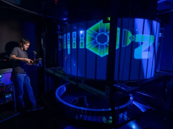
Behavior
Instantaneous Cue Rotation Arena
One of the behavioral apparatuses used for rodent spatial navigation tasks.
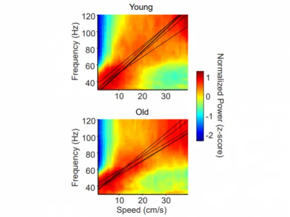
Electrophysiology
Electrophysiology in rodents
Electrophysiological recordings are used to investigate brain activity and electrical signals while animals perform tasks.
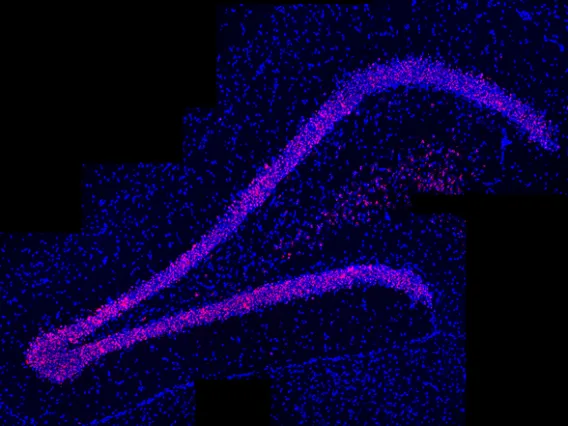
Molecular Biology
In situ Hybridization
Fluorescent in situ hybridization is used to tag and visualize mRNA expression in brain regions of interest.
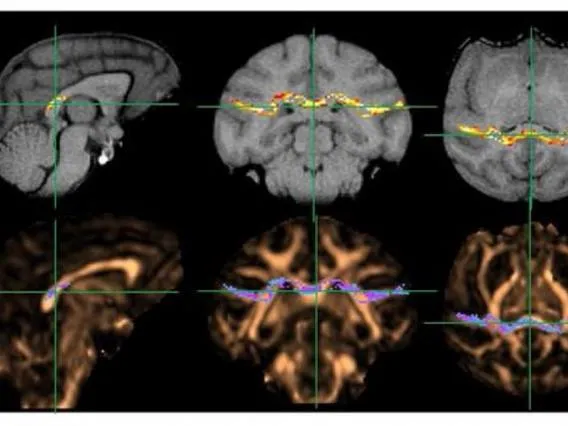
Anatomy: MRI
Magnetic Resonance Imaging (MRI)
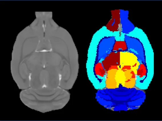
MR images are gathered and used to examine brain structures and study potential changes between young and aged animals.
Anatomy: IHC
Immunohistochemistry (IHC)
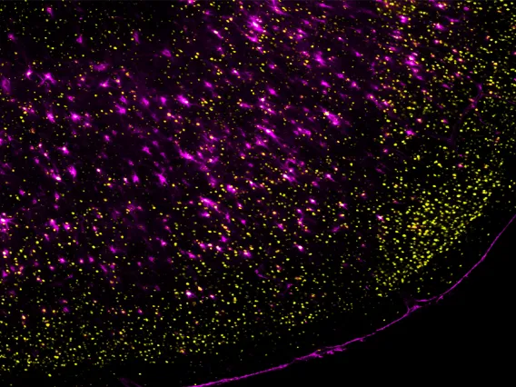
Using immunohistochemistry, fluorescent labeling, and microscopy we can visualize various neurobiological features in brain regions of interest.
Anatomy: MRI Mapping
MRI Processing
MR images are mapped onto labeled brain atlases to get anatomical specificity.

